UAMS Eye Patient: To See Again is Phenomenal
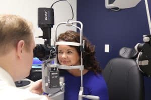
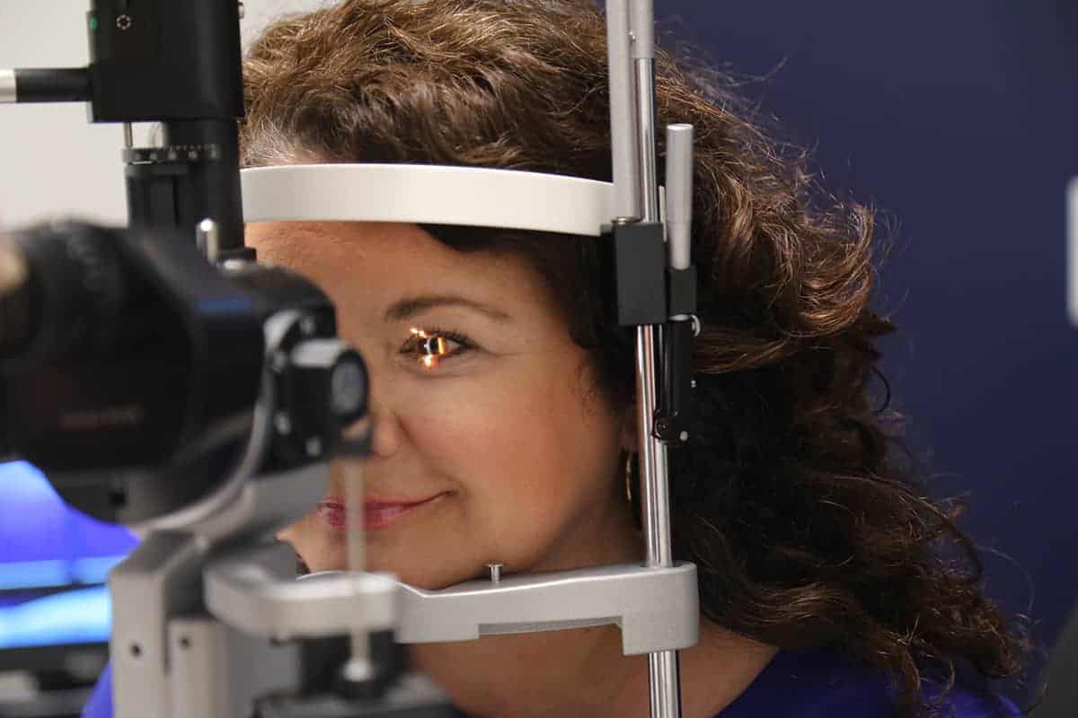
| June 25, 2018 | Each visit to UAMS to see cornea surgeon David Warner, M.D., is a joyous occasion for Cindy Jones.
That’s because with each visit in the year-long recovery after her cornea transplant, her vision improves.
“Each time it was better and better,” said Jones.
Jones, 44, has had a lifetime of vision problems. She was first prescribed glasses at 12 and was diagnosed with keratoconus at 14. The disease affects the cornea, progressively shifting and thinning the normally round cornea into a bulgy, cone-like shape. It can worsen until a person has difficulty seeing to perform daily activities.

Jones first came to Warner in 2016 to treat her keratoconus, an eye disease that affects the cornea.
“It’s a fairly common condition and a person usually falls into one of three categories with it,” said Warner. “Some have no progression and see well without glasses, others require hard contact lenses and avoid surgery. The third group usually has corneas that are so steep and bulgy it requires surgery.”
Jones tried glasses and contacts to no avail. She often squinted throughout the day to see better and relied on her daughter while driving at night to make sure she didn’t miss any turns.
It was an upcoming driver’s license renewal and eye screening in 2016 that brought the Cabot native to Warner, an ophthalmologist at the UAMS Harvey & Bernice Jones Eye Institute, assistant professor and director of the cornea service in the UAMS College of Medicine’s Department of Ophthalmology.
Warner said Jones would need corneal transplants in both eyes.
“She had a little different surgery than normal,” said Warner. “In one eye, we performed the traditional full thickness transplant, but the other eye received a more elegant approach, a partial thickness transplant.”
It removed about 95 percent of the cornea and allowed Jones to keep the corneal layer that prolongs the life of it, said Warner.

Since coming to JEI, Jones said she “can see things I had never noticed before.”
Both corneas came from the Arkansas Lions Eye Bank & Laboratory, housed in Jones Eye Institute.
“Because the eye bank is here, we have firsthand knowledge of how these tissues are recovered, evaluated and prepared for donation,” said Warner, the eye bank’s medical director. “It gives us great confidence that each patient receives an excellent graft.”
Recovery for a corneal transplant is about a year, said Warner.
“The grafts heal over the first six months, then we begin removing the sutures,” said Warner. “That happens two at a time until the astigmatism is almost gone or the sutures are gone, and we can correct the rest with contact lenses or glasses.”
Jones said she remembers being able to see better right away. The first few months required an eye patch and eye drops. Then, real change occurred with the first suture removals.
“I can see things I had never noticed before,” said Jones. “This is wonderful for me. I’m another one of Dr. Warner’s success story and I’m honored by that.”
The last of her sutures were removed in May. She will be fitted for glasses in June. Gone are Jones’ day of spotty vision.
“Being legally blind and then being able to see is phenomenal,” she said.
