First-Year Med Students Showcase Anatomy Findings
| Teams of first-year College of Medicine students concluded their nine-week full-body dissection course recently by presenting their findings before faculty and peers.
The Human Structure Student Research Conference was held Oct. 12 in the Education II building and featured 35 teams of five. The students were asked to prepare professional written and oral presentations detailing their dissection discoveries, including any possible causes of death.
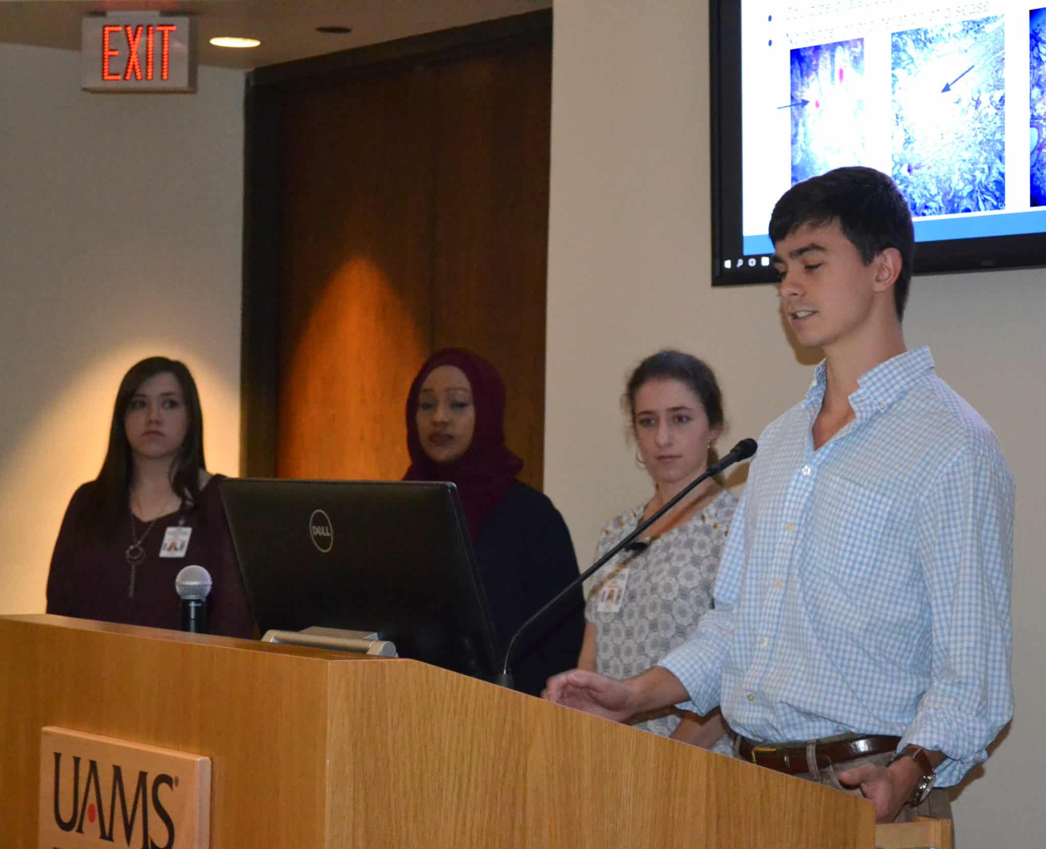
Wearing their short white coats and taking turns at the mic, each team led the group through slides detailing their experience with the dissection and any interesting anatomy. They discussed their hypotheses and detailed the research journey. Often, discoveries led them on the search for more information, which led to more discoveries.

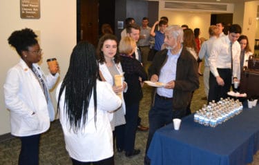
Students and teachers chat during one of the breaks during the conference. In total, 35 teams of five completed presentations.
“For most of us, this was our first experience like this. A lot happened, and the conference was a good way to look back on the journey as a whole,” said student Hytham Al-Hindi.
While dissections have long been a key part of the study of human anatomy during medical school, the course at UAMS is different in a couple of key ways. For one, students are given time to reflect, said David L. Davies, Ph.D., co-director of the Division of Clinical Anatomy in the College of Medicine.
“This is one of two end-of-course events for students,” Davies said. “The point of the conference is to consider the scientific aspects of their experience. It gives them a chance to revisit the learning objectives and look at the overall picture. After they return from fall break, we also hold a memorial service, which is a different sort of reflection on their emotional experience with the dissection.”
In addition, the course structure encourages the students to work in teams from the start of their medical careers.
Davies started the conference in 2014 with Charles Matthew Quick, M.D., an associate professor in the Department of Pathology in the College of Medicine. They successfully obtained grant funding from the Arkansas Medical Society for the conference and to add pathology to the students’ dissection experience in order to deepen its investigative qualities.
For instance, if the students came across something interesting during the dissection, they could take a biopsy and send it to the Department of Pathology’s clinical laboratory, where the tissue was processed and given to the teams in the form of microscopic slides for them to examine. This year, Rebecca Levy, M.D., played an especially significant role in this lab activity.
Another multidisciplinary aspect of the course comes from the Department of Radiology, which prepared CT scans of the dissection subjects for several of the groups. Sharp Malak, M.D., clinical co-director of the Human Structure module, met with about a dozen teams to review and guide interpretation of the CT scans, which were displayed on the Division of Clinical Anatomy’s new Sectra Table — a 4K resolution screen and software that behaves like a larger-sized version of a common smart phone. Students can swipe, scroll, zoom and rotate through the CT scans and explore anatomy in the process.
“We were truly working like a care team from the start,” said Monroe Albertson.
Al-Hindi, his team partner, agreed: “It was cool to integrate not just anatomy, but other disciplines like biology, radiology and pathology. They were all part of the experience from the start, so it wasn’t your typical dissection course.”
Davies hopes this approach results in more well-rounded learners — and future physicians.
“It builds their curiosity,” Davies said. “I don’t want them just memorizing. I want them thinking about what they’re doing and letting that interest drive their experience.”


