UAMS-Produced Head Molds to Improve Quality of Brain MRI
| How long do you think you could lie still without moving an inch? Five, 10, maybe 15 minutes?
A person undergoing a magnetic resonance imaging (MRI) scan is expected to lay still in a relatively small space for at least 30 minutes. Now, imagine having to go nearly twice that long without moving, without changing your position.
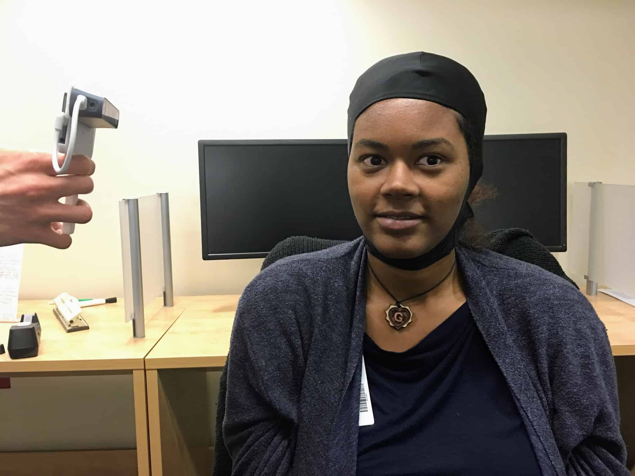
Guillory models the cap worn by research subjects as photos are taken with a cellphone.
Subjects in an MRI research study may be asked to spend between 45 minutes and an hour in an MRI scanner without making a move. Movement of the body, especially head motion, creates blurry images, which are difficult to interpret. Researchers in the UAMS Brain Imaging Research Center (BIRC), however, have a solution they believe will improve the quality of images produced during scans and, in turn, help them gain more insight into the puzzle that is the human brain.
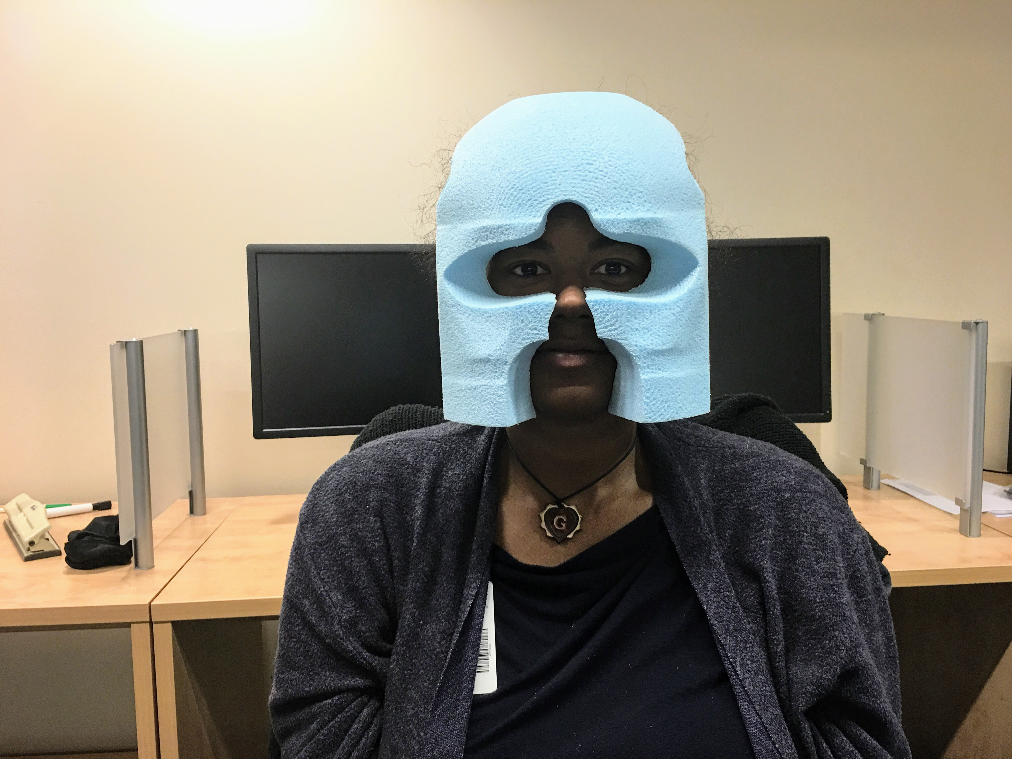
People taking part in research at the Brain Imaging Research Center now have the advantage of wearing a Styrofoam head mold while in the scanner. The molds are created to fit each individual perfectly and allow him or her to rest comfortably in the scanner with little to no movement of their head.
To create the head mold, a participant puts on a covering similar to a swim cap and sits still while the BIRC team takes photos using a cellphone with a special attachment for three-dimensional imaging. Photos are taken from 360-degrees around the subject to create a 3D model of his or her head. This model is transmitted via e-mail to a firm in California, which uses a device similar to a 3D printer to create a two-piece Styrofoam head mold that perfectly fits the subject’s head, complete with openings for the eyes, nose and mouth. The head mold is then shipped to the UAMS researchers within two to three days.
Now, thanks to a $359,000 award from the National Institute on Drug Abuse, UAMS is purchasing a device to produce the head molds, which will make the researchers the first in the world to produce these head molds on site.
Head motion by a subject while in the scanner is the biggest challenge researchers have for getting accurate brain imaging data, said Andrew James, Ph.D., an associate professor in the UAMS College of Medicine Department of Psychiatry. Using the head mold reduces motion dramatically and allows the researchers to get precise measurements of the subject’s brain activity as they perform certain tasks in the scanner.
“No one else in the world has this capability. We’ll be able take photos of a subject and produce a head mold within an hour,” said James. “This will speed up our imaging process and cut down on data loss from people who have trouble keeping their heads still in the scanner.”
The head molds will be particularly helpful in research related to adolescents and those with addiction issues, James said. Both groups have difficulty remaining still during MRI scans, more so than the average person.
“This will greatly improve our ability to acquire MRI data from those two groups,” he said. “Anything we can do to make them more still and comfortable is a big plus.”