View Larger Image
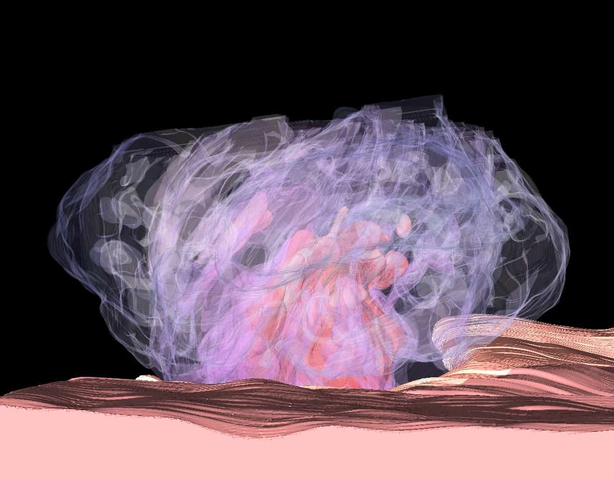
UAMS researchers Brian Storrie, Ph.D., and Irina Pokrovskaya, M.S., and colleagues at the National Institute of Biomedical Imaging and Bioengineering created this 3D rendering of a blood clot from a mouse, a winning entry in the 2019 BioArt competition.
Image by Brian Storrie/Irina Pokrovskaya
UAMS-Led Research Spurs Artistic Revelation
| It looks like something conjured by the imagination, or perhaps an other-worldly landscape. But an award-winning artistic image actually has roots in the laboratory and creative minds of researchers at the University of Arkansas for Medical Sciences (UAMS).
Brian Storrie, Ph.D., a professor in the UAMS College of Medicine Department of Physiology and Biophysics, and research associate Irina Pokrovskaya, M.S., used 3D renderings from sophisticated microscopic imaging techniques to create a winning entry in the Federation of American Societies for Experimental Biology (FASEB) 2019 BioArt competition.
Though it evokes a surreal world, the image actually shows a thrombus, or blood clot, that has formed inside the jugular vein of a mouse to close a puncture and stop bleeding. An aggregation of blood platelets appears as a wispy, pale purple cap over the reddish, globular mass of erythrocytes (red blood cells) within. Beneath the clot, the punctured jugular vein appears as a wavy pink and brown field. In the background, the lumen, or interior space inside the vein, appears black.
The UAMS researchers collaborated with Kenny Ling, B.S., Yajnesh Vedanaparti, B.S., Maria A. Aronova, Ph.D., and Richard D. Leapman, Ph.D., of the National Institute of Biomedical Imaging and Bioengineering (NIBIB) in Bethesda, Maryland, on the artistic project, and with additional collaborators at the NIBIB and the University of Kentucky in Lexington on studies that led to it.
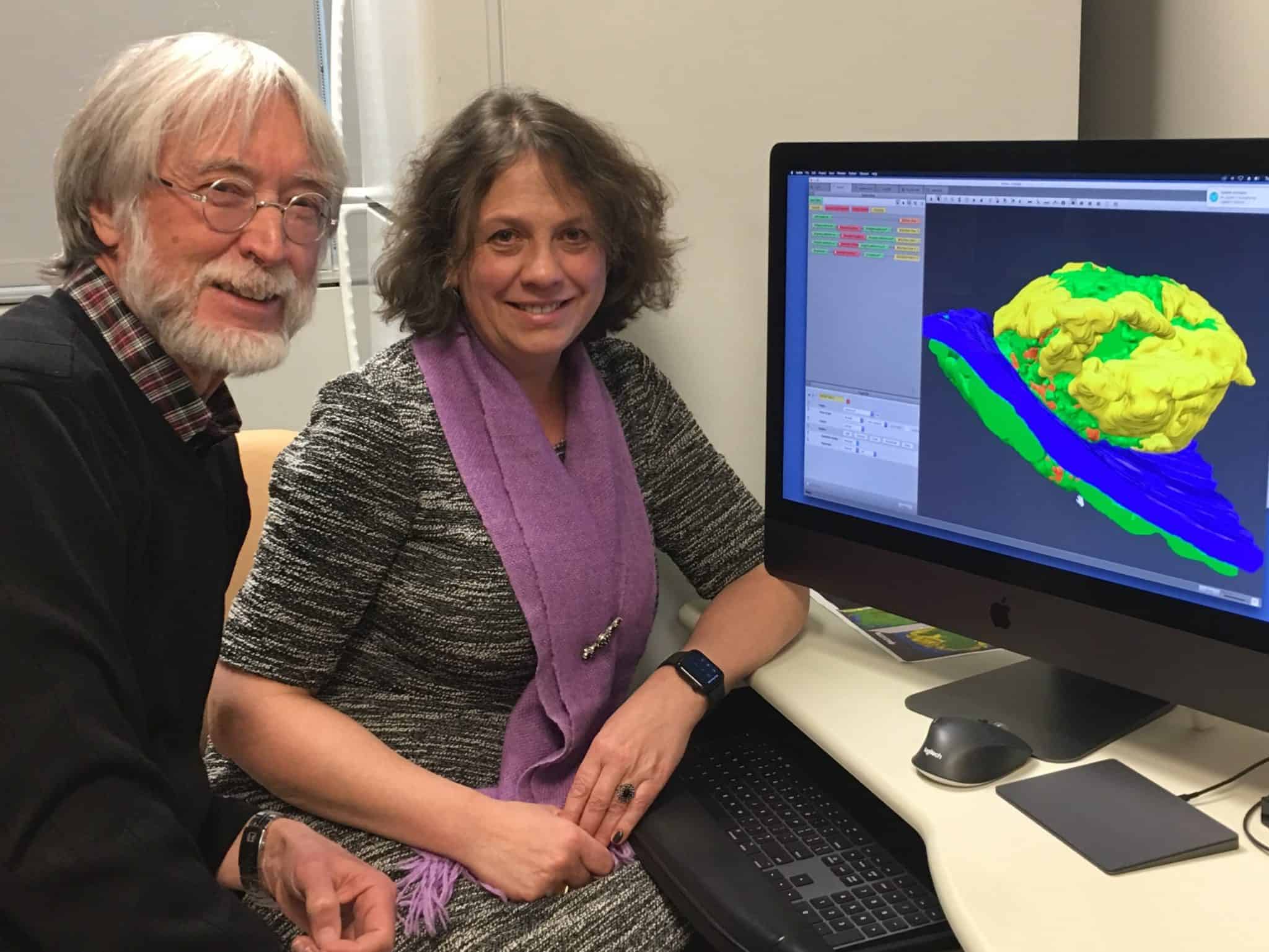
Brian Storrie, Ph.D., and Irina Pokrovskaya, M.S., view a 3D model of a thrombus on the computer.Tamara Robinson
“The actual image comes from playing with 3D renderings from 4,000 sequential cross section (images) of a five-minute post-wounding thrombus,” Storrie explained. “The cross-sections are spaced just 200 nanometers, or 0.2 microns, apart. Different layers of the structure can be made more or less transparent or translucent, and layers can be colored as wished.”
The genesis of the artwork was research into human platelet activation using ex vivo (outside of an organism) experiments. Storrie and colleagues at UAMS, the NIBIB and the University of Kentucky published two papers in the journal Blood Advances in 2018. The collaborators went on to consider what platelets do in vivo, or in the body, to form a thrombus to stop bleeding. The standard thinking had been that platelets form a plug that fills the hole of a wound, and the researchers began to explore this concept, working initially with mouse puncture wound samples produced by Tim Stalker, Ph.D., of the Perelman School of Medicine at the University of Pennsylvania in Philadelphia, and later with samples produced at UAMS.
At the NIBIB, the team produced 20 image sets, each consisting of thousands of micrographs across individual thrombi, using a serial block face imaging electron microscope and then rendered into 3D images. At UAMS, the team produced an additional 15 image sets using wide area transmission electron microscopy, stitching together hundreds of individual electron micrographs at selected depths within a thrombus to yield highly detailed subcellular imaging at a resolution of 3 nanometers.
Storrie and his colleagues are working to document their findings. An initial short article on platelet activation state was published in Blood in November 2019, and subsequent detailed manuscripts are being prepared to highlight bleeding cessation mechanisms, the activation state of individual platelets in a forming thrombus, and the structural organization of thrombi. Ultimately, their work could yield valuable information for potential therapeutic targets for preventing life-threatening blood clots.
In the process of pursuing scientific knowledge, the researchers found the opportunity for a creative diversion, and the result was the entry in the FASEB BioArt contest. Storrie emphasized that the NIBIB’s Kenny Ling and UAMS’ Irina Pokrovskaya also had lead roles in the endeavor.
“There is much satisfaction in realizing that what is true of us can be captured in a beautiful and striking image,” Storrie said. “This is a team effort, and my congratulations to the team.”