View Larger Image
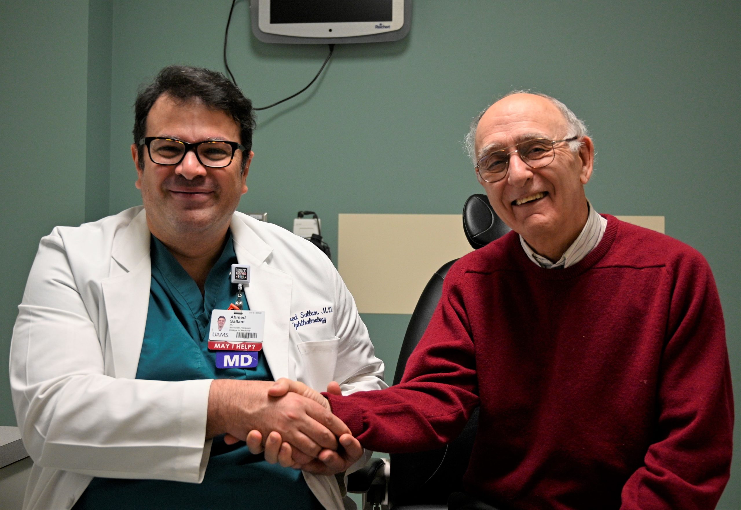
Ahmed Sallam, M.D., Ph.D., and patient Jim Winter.
Floater Removal Gets Patient Back on the Tennis Court
| Jim Winter realized something was wrong on the tennis court.
“In the last three to four years, I started having difficulty seeing tennis balls on the tennis court,” said Winter, a retired emeritus research professor at the University of Arkansas at Little Rock
Winter was also having trouble working on a computer or reading books or news print. “I worried about my quality of life decreasing.”
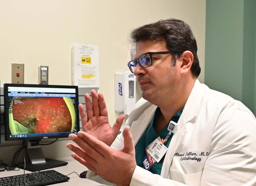
Sallam explains “floaters” with an image illustrating them in Winter’s right eye prior to surgery.
Winter had what are commonly called floaters building up in his right eye, turning the inside into something like a snow globe. In addition, he had cataracts and a small hole in the central part of the retina called the macula.
“You see specks floating around in your eye and when you do things like look up at the sky or a white wall, they really stand out. They move around in your vision,” said Winter.
“I was told there’s not much you can do about floaters and that it was too risky to treat them, so I decided to come to the UAMS Jones Eye Institute because I figured either someone here could help me or they would know who could. One of the JEI doctors told me he had difficulty seeing my retina because there were so many floaters, and here I was trying to see out through those floaters.”
That doctor referred winters to Ahmed Sallam, M.D., Ph.D., who specializes in vitreoretinal disorders and eye inflammation at the Jones Eye Institute.
“Floaters usually do not affect vision, so the usual advice is that if they’re not lowering your quality of life then leave them alone. But for Dr. Winter, they were,” said Sallam.
While surgery is usually a last option, in this case it was the only course of treatment. It involved removing the vitreous fluid of the eye to get rid of the floaters and replacing it with saline. Sallam also placed a bubble of gas to help Winter’s macular hole heal and inserted a new lens to treat the cataract.
“Having your eye operated on is always going to be a bit nerve wracking, but the procedure was really quite comfortable,” said Winter. “It only took an hour and there was no pain involved. Recovery was very quick. It took maybe two to three weeks.”
Sallam attributed the ease of the surgery to the technology involved.
“The level of technology we have here is fantastic. We operate using 3D surgery, which means that we operate not under a regular microscope but on a very big screen with augmented magnification and depth perception,” he said.
Rather than being hunched over the patient during an operation, the surgeon can see very fine details of the patient’s eye projected in 3D, which allows them to see the depth of the eye and layers of the retina as they work, increasing precision and patient safety.
“With the 3D surgery we have built-in live retinal imaging. As I operate, I see exactly what’s happening. As I peel a membrane off the retinal surface, I can see exactly what the retina looks like and how the tissues respond. It makes surgery very precise. There’s no guessing to what is happening.”

A picture of the interior of Winter’s eye shows no floaters after surgery.
This “head up” technique for surgery creates less strain on the surgeon’s back and neck and greatly helps in teaching students and residents, because the entire operating room can see exactly what the surgeon is doing. The operation can be recorded and watched again in 3D for teaching and training.
For all the technological complexity, however, the intent is to create minimal disruption for the patient.
“In surgery, the patient is awake, but we give them a numbing medicine around their eye. It’s a safe way of doing things. It decreases any disruption of the eye and relieves the need for general anesthesia,” said Sallam. “We use minimally invasive surgery with very tiny instruments that make very tiny holes in the eye that we don’t usually need to suture. We use local anesthesia with minimal or no narcotics or sedation, so there’s no disruption to the patient’s life. Nearly all our surgeries are outpatient surgeries, and patients can just go home afterward.”
Since his surgery in late 2019, Winter is indeed back home on 2.5 acres just outside Little Rock enjoying the native trees, flowers and walking his dogs – that is, when he’s not on the tennis court.
“The end result is my vision is really good. It’s very bright and I can see a wide, panoramic view. I’m so elated to have quality of life back.”