View Larger Image
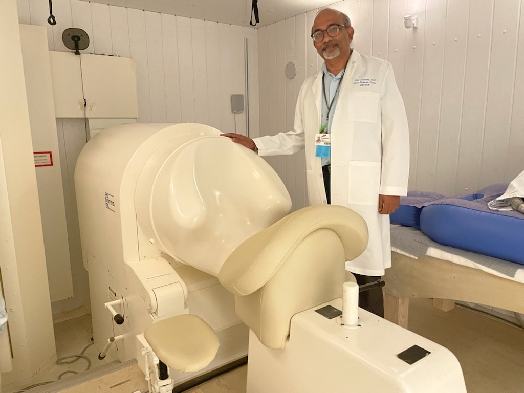
Hari Eswaran, Ph.D. ,shows off SARA, which he helped develop.
NIH Awards $1.36 million Grant to Aid UAMS Research on Pregnant Women’s Pelvic Floor Muscles
| The National Institutes of Health has awarded a $1.36 million grant to researchers at the University of Arkansas for Medical Sciences (UAMS) to develop a non-invasive means of detecting weaknesses in a pregnant woman’s pelvic floor muscles that could lead to injury while giving birth.
The principal investigators are Hari Eswaran, Ph.D., a professor who is the director of research for the College of Medicine’s Department of Obstetrics and Gynecology and the Institute for Digital Health & Innovation, and Sallie Oliphant, M.D., MSc, an assistant professor in the department who is board-certified in Obstetrics and Gynecology and Female Pelvic Medicine and Reconstructive Surgery.

Sallie Oliphant, M.D., MSc.
Over the past several years, Eswaran and Oliphant compiled preliminary data indicating that they could use a device originally designed to measure fetal brain and heart activity to better assess the strength and activity of a pregnant woman’s pelvic floor muscles before her delivery date. They then compared the functionality of the women’s muscles after childbirth to assess the effects of pregnancy and delivery on the pelvic floor.
“Maternal birth injury clearly contributes to the development of pelvic floor disorders, yet we possess an incomplete understanding of the impact of maternal pelvic support adaptations, birth injury and injury recovery on pelvic floor support,” Eswaran said. “Using the novel approach of magnetomyography, our project will combine clinical, anatomic and physiologic endpoints to improve our knowledge of maternal pelvic muscular adaptations and injury recovery patterns.”
Magnetomyography is a technique for mapping muscle activity by recording magnetic fields produced by electrical currents in the muscles.
“From a clinical standpoint, we hope our work will shed light on the impact of pregnancy and childbirth on the pelvic floor support muscles,” said Oliphant. “We know that these muscles are structurally critical to pelvic health and that injury to these muscles is associated with conditions such as urinary and fecal incontinence and pelvic organ prolapse. We hope to use magnetomyography to better understand how these important muscles are altered by the birth experience.”
The device that will be used in the four-year study to develop analysis tools to enhance assessment capabilities is known as SARA (SQUID-Array for Reproductive Assessment) and was developed at UAMS in 2000 by a team that included Eswaran. SQUID stands for Superconducting Quantum Interference Device.
SARA is equipped with 151 sensors cooled in liquid helium. For more than 20 years, it has been used to obtain three-dimensional data from the fetus and the uterus, without the use of any invasive instruments, when a pregnant woman sits leaning forward against a concave shield that covers her abdomen.
The research, which includes a dedicated research nurse, Heather Moody, RN, has since found that when a pregnant woman sits on the machine facing the opposite direction and performs Kegel exercises as directed by a technician whose voice is piped into the closed room, doctors can “see” eight centimeters deep into the pelvis – much further than they could before – and better assess her pelvic floor muscles.
The pelvic floor muscles are located deep in the pelvis. They support the uterus, bladder, small intestine and rectum and can become weak through pregnancy, childbirth and straining due to constipation or other events, including improper exercises. Before magnetomyography, the ability to non-invasively assess any potential weakness in the muscles has been limited to interpreting electrical activity that is recorded by surface electrodes placed on the skin above the muscles.
Kegel exercises involve repeatedly contracting and relaxing the muscles of the pelvic floor, which are collectively known as the levator ani muscles. The enhanced information available by using SARA also allows technicians to determine whether the woman is properly squeezing only her Kegel muscles and if not, to guide her to isolate them.
UAMS is the state’s only health sciences university, with colleges of Medicine, Nursing, Pharmacy, Health Professions and Public Health; a graduate school; a hospital; a main campus in Little Rock; a Northwest Arkansas regional campus in Fayetteville; a statewide network of regional campuses; and seven institutes: the Winthrop P. Rockefeller Cancer Institute, Jackson T. Stephens Spine & Neurosciences Institute, Harvey & Bernice Jones Eye Institute, Psychiatric Research Institute, Donald W. Reynolds Institute on Aging, Translational Research Institute, and the Institute for Community Health Innovation. UAMS includes UAMS Health, a statewide health system that encompasses all of UAMS’ clinical enterprise. UAMS is the only adult Level 1 trauma center in the state. UAMS has 3,553 students and 1,030 medical residents and fellows, and two dental residents. It is the state’s largest public employer with about 12,000 employees, including 1,200 physicians who provide care to patients at UAMS, its regional campuses, Arkansas Children’s, the VA Medical Center and Baptist Health. Visit www.uams.edu or uamshealth.com. Find us on Facebook, X (formerly Twitter), YouTube or Instagram.###