UAMS Research Team Upends Understanding of How Blood Clots Form; NIH Awards $2.5 Million for Further Study
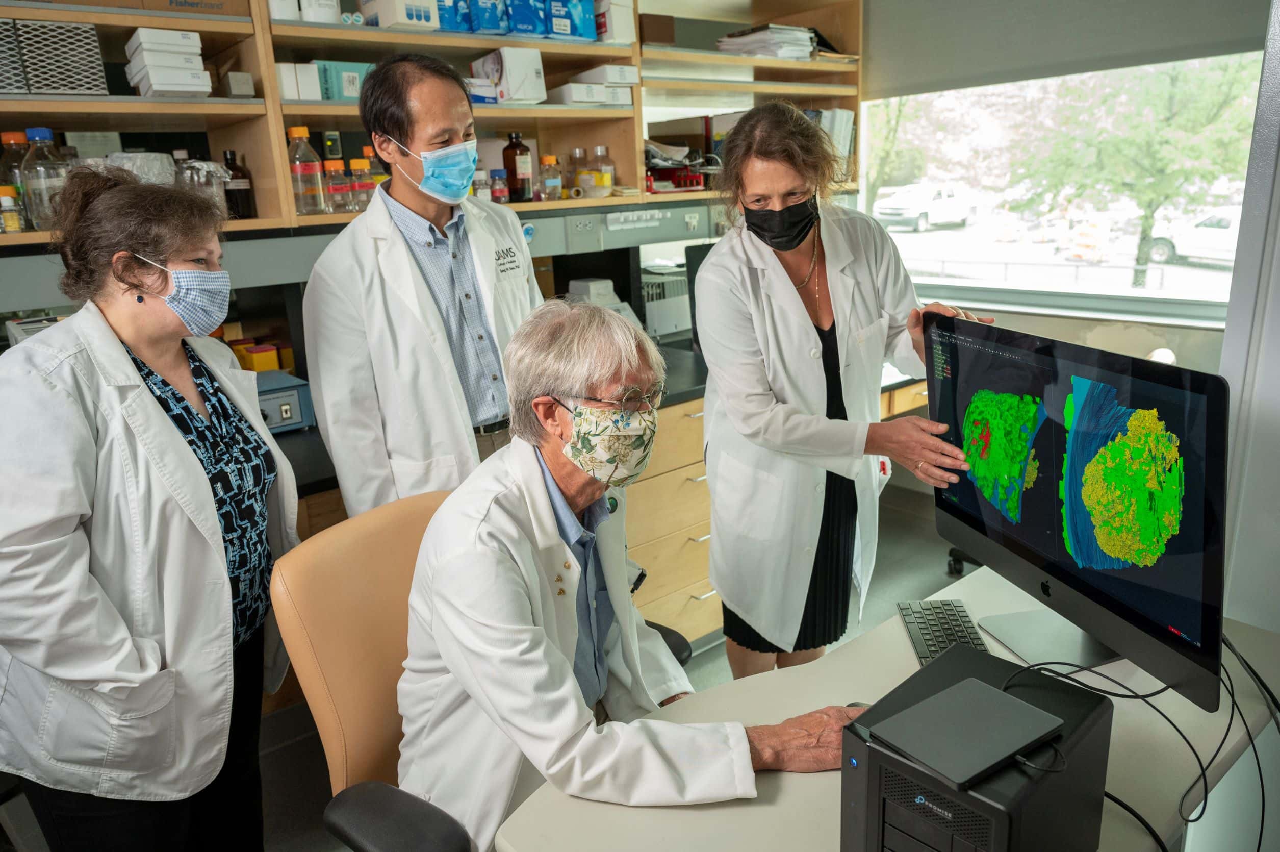
| LITTLE ROCK — A UAMS-led research team has found that blood clots form in puncture wounds similar to a skyscraper, with rooms and furnishings that scientists can now see. Published in Communications Biology, the discovery of the vaulted thrombus (blood clot) structure surprised researchers and is a big change from a long-held hypothesis.
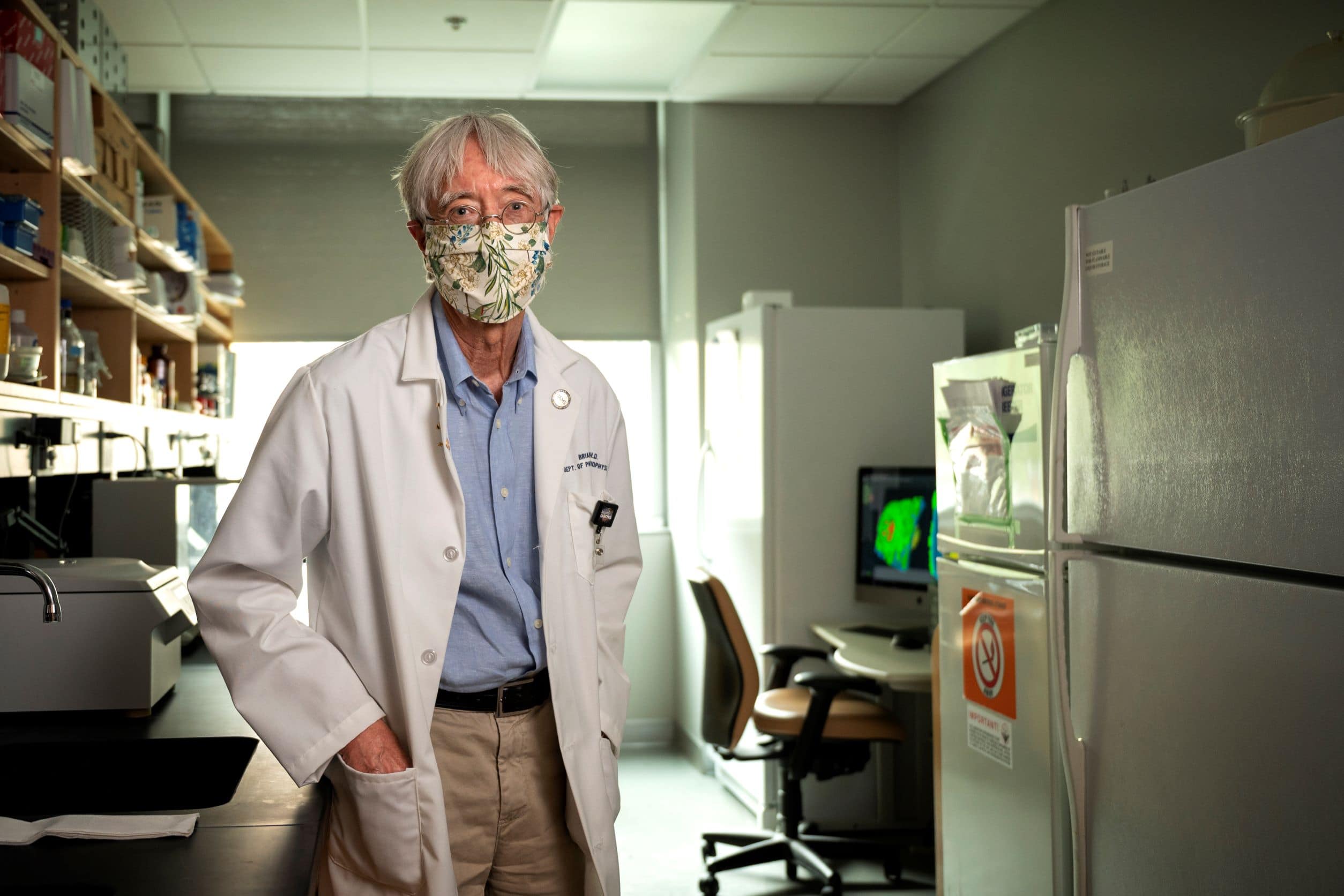
Brian Storrie, Ph.D., is the project’s principal investigator.
The Sept. 16 Communications Biology publication follows a $2.5 million National Institutes of Health (NIH) award in August to expand on the research by UAMS’ Brian Storrie, Ph.D., lead author and principal investigator. The findings could affect drug development for bleeding regulation, which is one area he will explore as part of the grant.
Collaborators outside of UAMS include researchers from the University of Kentucky and the NIH National Institute of Biomedical Imaging and Bioengineering (NIBIB).
Keys to the research are advanced fluorescence microscopy techniques and 3D electron microscopy, which provides high-resolution images of the clot formation.
“We got to know the whole structure of the thrombus by taking thousands of electron micrographs,” said Storrie, a professor in the College of Medicine Department of Physiology and Cell Biology. “We know exactly where all the rooms are in the skyscraper and how the furniture is arranged.”
“The research by Dr. Storrie and his team provides a fascinating image at an incredibly detailed level into how blood clots form and vary in different tissues. This insight should help pave the way for future drugs to prevent life-threatening blood clots while minimizing the risk of bleeding,” said Susan Smyth, M.D., Ph.D., executive vice chancellor of UAMS and dean of the College of Medicine, a cardiologist whose own research and clinical care has centered on prevention and treatment of thrombosis.
The leading hypothesis for how the body stops bleeding in a puncture wound has been a “core and shell model,” in which platelets (colorless blood cells that help blood clot) fill the hole layer by layer, forming a plug in the wound.
“Our results are very different than what has been thought to be true of blood clot formation,” Storrie said.
His research points to a “cap and build” concept, with the formation of pedestals and columns that merge and are capped by platelets. The “rooms” in the skyscraper-like structure are spaces that are compressed in later phases of clot development.
The research team’s state-of-the-art approaches reveal the activity of single platelets in blood clots. Storrie said the findings are unequaled in demonstrating the spatial and time resolution of clot formation.
While significant, the team considers its findings a first step in the structural understanding of bleeding cessation.
Over the next four years, Storrie and his team will use the NIH R01 grant to study molecular-level coagulation factors and signaling between platelets that occurs immediately before and during the clot formation. Their work will include analyses of common anticoagulant drugs such as cangrelor and dabigatran and how they affect the signaling in thrombus formation.
They will also test hypotheses that developed in their earlier research.
UAMS researchers on the project are: Sung W. Rhee, Ph.D. (co-principal investigator); Jerry Ware, Ph.D.; Irina D. Pokrovskaya, M.S.; Kelly K. Ball, M.S.; and Jeffrey A. Kamykowski.
The team also includes Sidney W. Whiteheart, Ph.D., from the University of Kentucky and researchers from the NIBIB Laboratory of Cellular Imaging and Macromolecular Biophysics: Maria A. Aronova, Ph.D.; Richard D. Leapman, Ph.D.; Kenny Ling; Yajnesh Vedanaparti; Joshua Cohen; Denzel R. D. Cruz; Guofeng Zhang, Ph.D.; and Oliver S. Zhao.