View Larger Image
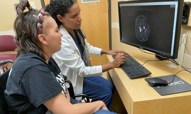
Analiz Rodriguez, M.D., shows Ashley James an image of her brain.
UAMS Neurosurgeon Uses Brain-Mapping Software, Laser Ablation to Safely Destroy Mom’s Brain Tumor
| | Just three months after giving birth to her daughter, Ashley James learned that a brain tumor she’d had removed nearly two years earlier had returned.
Back in October 2020 during the height of the COVID-19 pandemic, the Pine Bluff native was living in Cleveland, Ohio, when she experienced a severe headache and pain behind her eyes that wouldn’t go away. A trip to the emergency room for what she thought was a sinus infection resulted in the discovery, through an MRI, of a brain tumor. A biopsy confirmed that the mass was a glioblastoma multiforme (GBM), a fast-growing tumor in her right frontal lobe.
About 900 miles away in Arkansas, James’ mother and sister boarded a plane for Cleveland as she prepared for major surgery — a craniotomy — two weeks later to remove the cancerous growth.
The surgery successfully removed the tumor, but during it James suffered a seizure that left her unable to walk or to move the fingers on her left hand when she awoke. Doctors say this can happen during brain surgery when functionally critical nervous tissue is inadvertently removed along with diseased tissue because the intertwined matter cannot be distinguished through imaging techniques.
After months of physical therapy, James was able to walk and use the left side of her body again. She also underwent regular MRIs to check for any regrowth of the tumor. But when she became pregnant with her daughter — her third child — she was forced to stop the MRIs for several months because the dye used in the procedure posed potential health risks to her unborn child.
When her daughter was 3 months old and the MRIs resumed, she was devastated to learn July 8 that the tumor had reappeared, this time in three places: next to the original site and in two new spots in her brain.
Now 35 and living back in Arkansas to be close to family, James sought the expertise of neurosurgeons at the University of Arkansas for Medical Sciences (UAMS).
When she learned that laser ablation was available at UAMS, “I was really excited because it was less invasive, and I was terrified to have another craniotomy,” James said.
In the minimally invasive procedure, which James had heard about through an online gliobastoma multiforme support group but knew wasn’t yet widely available, a thin laser probe is inserted into the brain and used to blast away cancerous cells with heat.
UAMS is the only hospital in Arkansas that offers the treatment.
James was also thrilled when Analiz Rodriguez, M.D., Ph.D., a neurosurgeon at UAMS, offered to perform the first-ever procedure combining laser ablation and a revolutionary new brain-mapping technology called Quicktome.
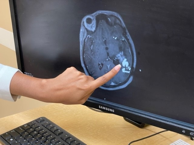
Rodriguez points to the area of James’ brain where the tumor was.
James hadn’t heard about brain-mapping technology, but was intrigued when Rodriguez described it. It enables surgeons to visualize patients’ brain pathways from their computer screens and map out the danger zones before surgery, in order to steer clear of them.
“We need to know where these critical brain pathways are located because we don’t want to enter them or work near them because then that could cause harm to the person, and they could wake up with problems talking, walking or seeing,” said Rodriguez, M.D., who is also the director of Neurosurgical Oncology at UAMS.
Brain networks are responsible for everything from language to movement to thought. While previous imaging solutions helped surgeons avoid the networks associated with major disability, many patients still emerged with cognitive, emotional and behavioral complications. Quicktome, approved by the FDA in 2021, is the first platform that allows mapping of the brain’s cognitive and emotional regulation networks.
In preparing for James’ Aug. 25 surgery, which Omniscient Neurotechnology confirmed was the first time that its Quicktome platform was used in conjunction with laser ablation, “our goal was to ensure the left side didn’t get weak again,” Rodriguez said. “Ashley had worked really hard through physical therapy so she could walk again, and her baby was only 3 months old.”
Rodriguez said part of the tumor was located near the area of James’ brain that controls the motor pathways, “so there was a risk that the ablation would cause left-side weakness or the inability to move for an extended period — or even permanently. Quicktome allowed us to map the motor pathways to know exactly what we were heating, to avoid causing any harm.”
She said a second craniotomy, in which the skull is opened to access the brain, isn’t recommended for recurring brain tumors, especially those reappearing in new locations, and would require a much longer recovery time.
Rodriguez said that after a biopsy that took about 45 minutes, the Quicktome-guided laser ablation surgery lasted two or three hours. When James awoke, she was relieved to find that she could move and feel all her limbs.
“I was able to go home the next day and take care of my baby and continue with physical therapy,” James said. “And honestly, with the help of steroids I took after brain surgery, I felt like Superman.”
In addition to Hadley Sue, James has two sons — 13-year-old Kadon and 15-year-old Landon.
“I have two older boys, and I wanted a girl so bad,” she said. “I was worried that I probably wouldn’t be able to have babies after the chemo, but God blessed me with a little girl.”
As a single parent, James said she didn’t know how she would have managed to take care of a newborn if the brain surgery had left her disabled, despite the help of a cousin she lives with.
She is now in the process of returning to UAMS five days a week through mid-November for chemotherapy and radiation treatments to ensure the tumor is gone, and then will return for increasingly less-frequent chemotherapy maintenance sessions.
“I feel pretty blessed,” James said. “I’m not a rocket scientist, but I know I’ve got a great team of brain scientists taking care of me. In fact, when I was first diagnosed, I thought GBM stood for Great Big Monster, but after all I’ve experienced, I know it means Great Big Miracle.”