Research Collaboration Crosses Basic Science, Clinical Boundaries
| Biomedical research does not exist in a vacuum. To succeed, scientists must work together by combining resources and ideas in new and innovative ways.
Many times, these collaborations exist across institutions, where combined strengths provide the perfect combination of skills necessary to undertake complex projects successfully.
Such is the case with a partnership between the UAMS Winthrop P. Rockefeller Cancer Institute and the University of Arkansas, Fayetteville (UA) that recently resulted in a five-year, $2.03 million R01 grant from the National Cancer Institute (NCI).
It all started with grant recipient Narasimhan Rajaram, Ph.D., and his mission to create a method of monitoring a cancer patient’s response to radiation and chemotherapy, allowing for timely changes to the treatment plan if necessary. Rajaram is a UA assistant professor of biomedical engineering.
With conventional treatment and monitoring methods, doctors are unable to determine how well a tumor responds to treatment until after the fact. This includes whether the patient’s tumor completely disappears, shrinks, remains the same or enlarges.
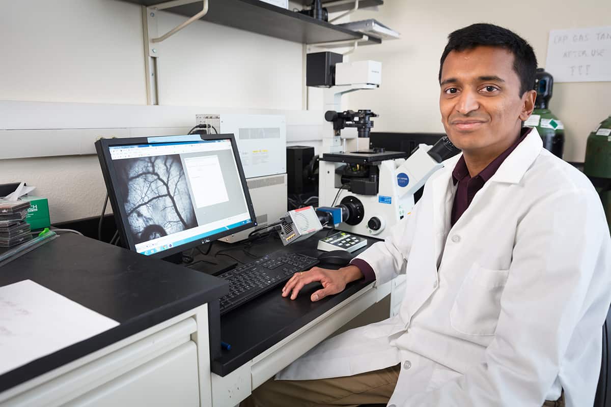
Grant recipient Narasimhan Rajaram, Ph.D., at work in his lab at the University of Arkansas College of Engineering.University of Arkansas
“Unfortunately, there are currently no methods that can identify treatment response in the clinic during therapy, which causes patients – both responsive and resistant – to lose critical time when alternative approaches could be considered,” said Rajaram.
To complete his experiments, Rajaram, who spent four years at Duke University before arriving at UA in 2014, required unique cell lines to examine for potential new biomarkers and disease-resistance patterns.
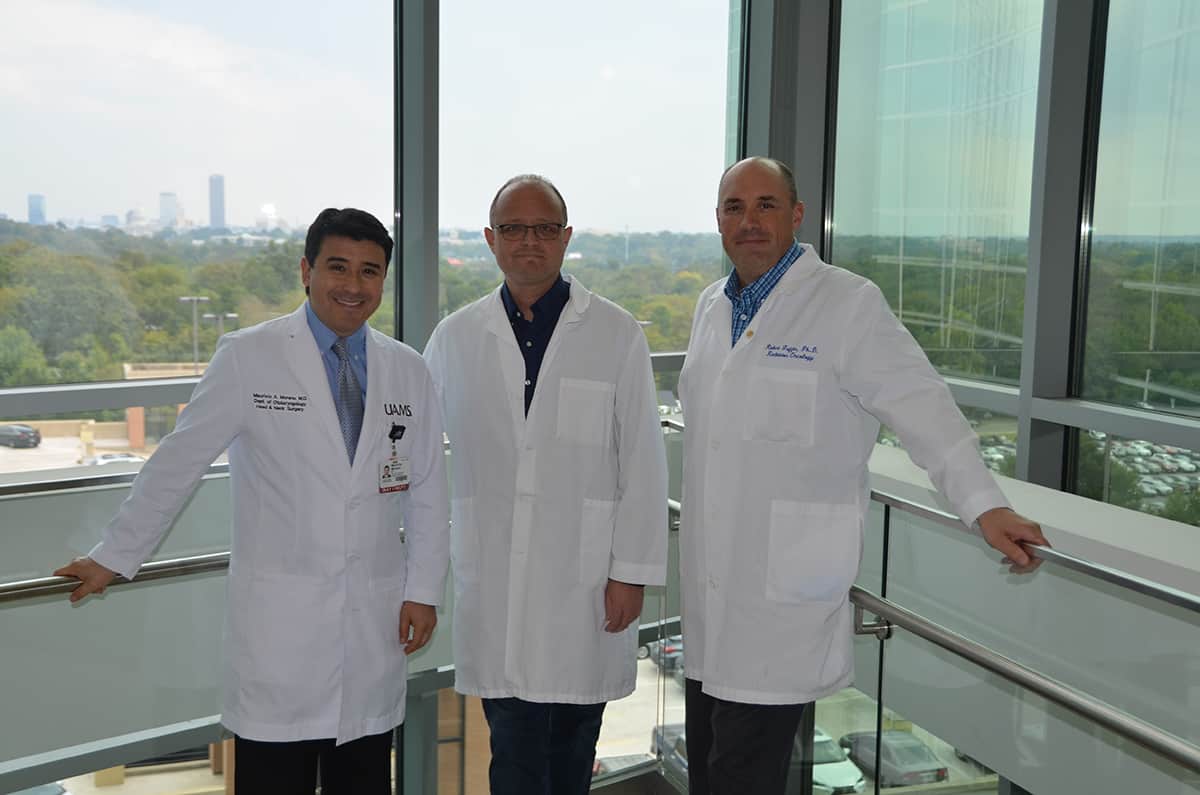
Through his network of colleagues, Rajaram discovered a connection to Robert J. Griffin, Ph.D., professor and biologist in the UAMS College of Medicine Department of Radiation Oncology. After initial discussions, the two determined their mutual interest in the effects of radiation therapy and disease resistance could lead to a partnership.
Griffin’s colleague Ruud Dings, Ph.D., who joined UAMS during this time, also entered the discussion. Dings is assistant professor in the Department of Radiation Oncology and, like Griffin, uses his laboratory to create unique cell lines.
“Previously, scientists studied disease resistance patterns by comparing cell lines that were not related to each other or that had different backgrounds, resulting in potential false correlations,” said Dings.
The unique cell lines created in the UAMS labs are isogenically related, meaning they are engineered from a parental line, have the same genetic background and are exposed to repeated interventions that create acquired resistance to radiation or drugs.
With a set of unique cell lines he acquired from Dings’ lab in 2015, Rajaram continued his work on optical imaging technologies, with the goal of developing a method to predict a tumor’s sensitivity to treatment.
A paper highlighting findings related to this research was published in the April 17, 2019, issue of Cancer Research, a publication of the American Association for Cancer Research.
Out of Rajaram’s research came the development of a thin, chopstick-like probe. When touched to a tumor or surrounding tissue, light emitted from the probe refracts. The light is then captured back into the probe and collected for analysis.
With the probe ready to test in humans, Rajaram reached out to UAMS surgeon Mauricio Moreno, M.D., for input on how to apply this emerging technology to head and neck cancer.
From there, the idea for a clinical trial was born.
“The Holy Grail of head and neck oncology is the ability to accurately predict which patients will or will not respond to radiation therapy,” said Moreno, associate professor in the UAMS College of Medicine Department of Otolaryngology-Head and Neck Surgery.
Those patients whose tumors do not completely disappear following radiation therapy typically require surgery. And, as Moreno pointed out, patients who have recently undergone radiation therapy are a much higher risk of potential surgical complications.
“If there was a way for us to know upfront that a patient was not likely to respond to radiation, we could alter their treatment plan and prevent them from experiencing the toxicity of radiation therapy in the first place,” Moreno said.
Moreno and Rajaram determined that head and neck squamous cell carcinoma (HNSSC) of the larynx or tonsil was the best condition on which to focus their clinical trial for four reasons:
1) It is a common cancer, resulting in a large number of potential participants.
2) It is treated with radiation.
3) The tumor can be easily accessed noninvasively.
4) It is often caused by the human papilloma virus (HPV), which is typically sensitive to radiation therapy.
Their study, which has enrolled about five UAMS patients so far, is approved to enroll up to 90 subjects with Stage 3 or Stage 4 HNSCC, including those with tumors that are both HPV-positive and HPV-negative.
Trial participants undergo both a baseline test after diagnosis and a test during their course of radiation treatment. The test includes inserting the thin probe into the patient’s throat and touching the probe’s light to the tumor and surrounding tissue. Researchers hope that by examining the light patterns captured during both of these tests they can identify correlations related to tumor treatment response.
“It’s very investigational, but if we can identify a pattern either before or during the early phases of treatment where we see the tumor is not responding properly, that could potentially have significant clinical impact for future patients,” Moreno said.
All data collected by the probe goes back to Rajaram for analysis and is compared to the patients’ other imaging results, including CT scans, for potential correlations.
The fact that Rajaram’s NCI grant funds both the basic science and clinical trial associated with this research is uncommon, said Griffin, adding that “the clinical observations collected by Dr. Moreno will strengthen the basic science conducted by Dr. Rajaram, Dr. Dings and myself.”
Additional pre-clinical studies and technology development were conducted at Johns Hopkins University. Rajaram’s initial work to demonstrate the feasibility of this approach was supported by start-up funds from UA and the Arkansas Biosciences Institute.
Research collaborations such as this one bolster the Cancer Institute’s ongoing efforts to receive National Cancer Institute Designation.
To achieve designation, cancer centers undergo a highly competitive assessment process that demonstrates an outstanding depth and breadth of research in three areas: basic laboratory, patient/clinical and population-based. The designation brings with it many benefits, including expanded access to federal funding for researchers and improved access to clinical trials for patients.